


Neuroendocrine Bronchial and Thymic Tumor
Get informed about Neuroendocrine Bronchial and Thymic Tumors with our expert reviews, patient care strategies, and latest research updates.

Many tests are used by doctors to detect or diagnose soft tissue sarcoma. They also perform tests to see if cancer has migrated to other parts of the body from where it began. It's known as metastasis when this happens. Imaging examinations, for example, can reveal whether cancer has spread. Images of the inside of the body are produced via imaging tests. Doctors may also conduct tests to determine which treatments are most effective.
A biopsy is the only guaranteed way for a doctor to know if a part of the body contains cancer in most types. A biopsy is a procedure in which a doctor removes a small sample of tissue to be tested in a laboratory. If a biopsy is impossible, the doctor may recommend further tests to aid the diagnosis. Although there is a slight chance that biopsies will not provide a definitive answer, they are critical in allowing your doctor to make a precise diagnosis and develop a team-based treatment strategy.
Also Read: What is Sarcoma?
This section discusses sarcoma diagnosis options. Not every person will be subjected to all of the tests described below. When choosing a diagnostic test, your doctor may take into account the following factors:
Sarcoma does not have any routine screening tests. Any strange or new lumps or bumps forming should be examined by a doctor to ensure they are not cancerous. If sarcoma is suspected, it is critical to consult with a physician who is familiar with this type of cancer.
A doctor's clinical examination and imaging tests diagnose a sarcoma. The results of a biopsy back this up. Some of the tests listed below, besides a physical examination, may be used to diagnose sarcoma.
Also Read: Treatment of Soft Tissue Sarcoma
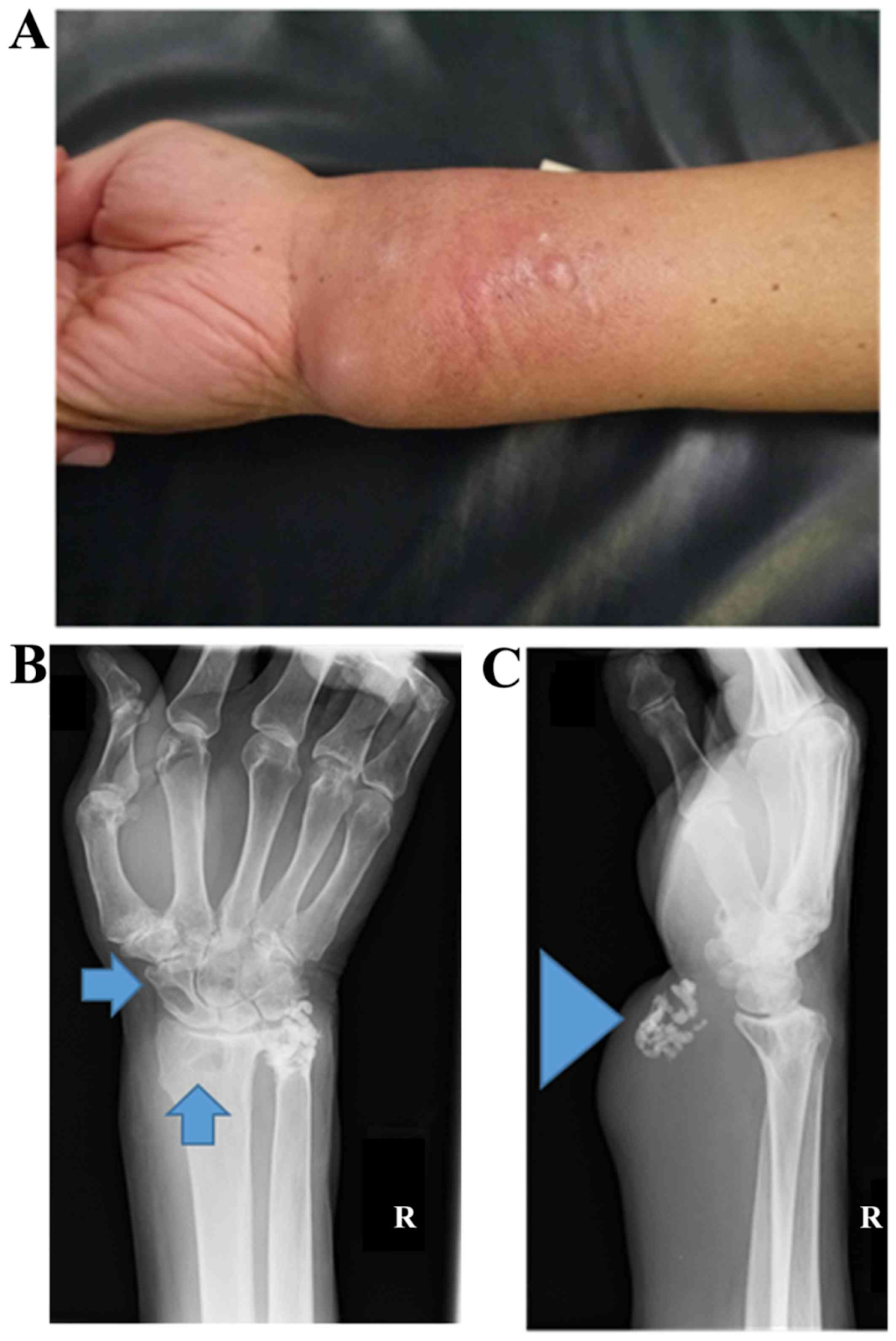
Imaging examinations, such as an X-ray, can detect benign and malignant tumours. A radiologist, a physician who performs and analyses imaging tests to identify disease, will use the appearance of the tumour on the test to assess whether it is benign or cancerous. A biopsy, on the other hand, is almost always required.
An X-ray uses a small amount of radiation to provide a picture of the structures inside the body. X-rays are very beneficial in the diagnosis of bone sarcomas, although they are less useful in the diagnosis of soft tissue sarcoma.
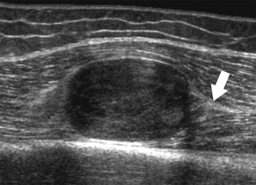
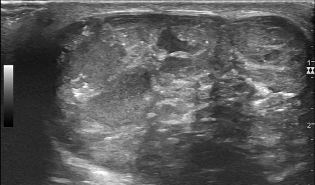
An ultrasound creates an image using sound waves and can be used to examine tumours under the skin or other organs in the body.
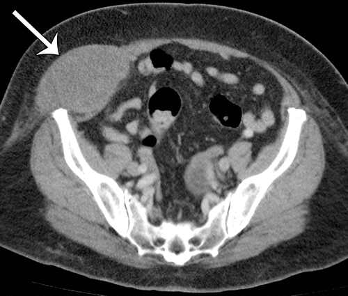
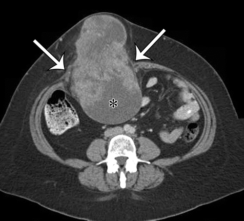
A CT scan uses X-rays captured from various angles to create images of the inside of the body. These images are combined by a computer into a detailed, three-dimensional image that reveals any anomalies or malignancies. A CT scan can be used to determine the size of the tumour or to determine if cancer has spread to other parts of the body. Before the scan, a dye called a contrast medium is sometimes used to improve image detail. This dye can be injected into a patient's vein or given to them as a tablet or liquid to swallow.
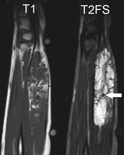
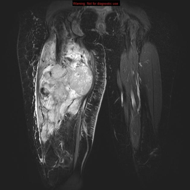
Magnetic fields, not X-rays, are used in an MRI to provide detailed body images. A magnetic resonance imaging (MRI) scan can be used to determine the tumour size. Before the scan, a dye called a contrast medium is administered to create a crisper image. A patient's vein can be injected with this dye. An MRI scan is frequently used to determine whether a sarcoma may be surgically removed.
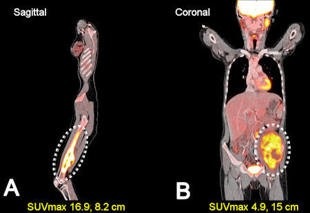
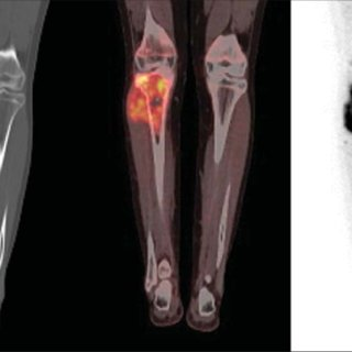
PET scans are frequently paired with CT scans (see above), resulting in a PET-CT scan. The patient is given a small amount of radioactive sugar to inject into his or her body. The cells that use the most energy absorb this sugar molecule. Cancer absorbs more of the radioactive substance since it uses energy actively. The material is then detected by a scanner, which produces images of the inside of the body. This technique can examine the tumour's shape and how much energy the tumour and normal tissues consume. This information can help plan treatment and assess how well it works, but it is rarely used in all cases of soft tissue sarcoma, whether known or suspected.
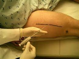
Although imaging tests may imply sarcoma, a biopsy is required to confirm the diagnosis and determine the type of sarcoma. Because a poorly performed biopsy might make surgery more complex, a patient must see a sarcoma specialist before undergoing surgery or a biopsy if a sarcoma is suspected.
A biopsy is a procedure in which a small piece of tissue is removed and examined under a microscope. Other tests may indicate the presence of cancer, but only a biopsy can provide a definitive diagnosis. A pathologist is a clinician who specializes in diagnosing disease by interpreting laboratory tests and assessing cells, tissues, and organs.
Because soft tissue sarcoma is an uncommon sarcoma, the biopsy should be reviewed by an experienced pathologist. Special testing on tumour tissue may be required to diagnose a sarcoma correctly, and it is preferable if this is done by a specialist who sees this type of cancer regularly.
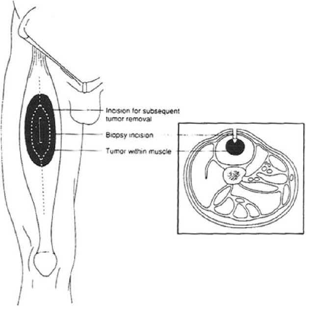
Biopsies come in a variety of forms.
When diagnosing and treating sarcomas, the type of biopsy and how it is performed are critical. Before the biopsy, patients should be evaluated in a sarcoma speciality facility so that the treating surgeon may choose the best place for the biopsy. To correctly identify a sarcoma, it is critical to have a pathologist analyze the tissue sample extracted.
The doctor or the pathologist who is examining the sarcoma may suggest that laboratory tests be performed on a tumour sample to identify specific genes, proteins, and other components that are specific to the tumour. Because each sarcoma is as diverse as breast and colon cancer, the results of these tests will aid in determining the best course of treatment.
Your doctor will review the results with you after the diagnostic tests are completed. These data can assist the doctor in describing cancer if the diagnosis is cancer. This is referred to as "staging and grading."
Enhance Your Journey with Positivity & Willpower
For personalized guidance on cancer treatments and complementary therapies, consult our experts at ZenOnco.io or call +91 9930709000
Reference:
Vodanovich DA, M Choong PF. Soft-tissue Sarcomas. Indian J Orthop. 2018 Jan-Feb;52(1):35-44. doi: 10.4103/ortho.IJOrtho_220_17. PMID: 29416168; PMCID: PMC5791230.
Vibhakar AM, Cassels JA, Botchu R, Rennie WJ, Shah A. Imaging update on soft tissue sarcoma. J Clin Orthop Trauma. 2021 Aug 20;22:101568. doi: 10.1016/j.jcot.2021.101568. PMID: 34567971; PMCID: PMC8449057.
 Link Copied


Nurture Hope & Healing
with ZenOnco
On Google play India


Nurture Hope & Healing
with ZenOnco
On Google play India


