


Retinoblastoma
Understand Retinoblastoma and how to manage it with our latest research, treatment strategies, and support services for families.

Many tests are used by doctors to detect or diagnose cancer. They also perform tests to see if cancer has migrated to other parts of the body from where it began. It's known as metastasis when this happens. Imaging examinations, for example, can reveal whether cancer has spread. Images of the inside of the body are produced via imaging tests of adenoid cystic carcinoma. Doctors may also conduct tests to determine which treatments are most effective.
A biopsy is the only guaranteed way for a doctor to know if a part of the body contains cancer in most types of cancer. A biopsy is a procedure in which a doctor removes a small sample of tissue to be tested in a laboratory. If a biopsy is not possible, the doctor may recommend further tests to aid in the diagnosis.
Also Read: Symptoms of Adenoid Cystic Carcinoma
The options for diagnosing adenoid cystic carcinoma are described in this section. Not every person will be subjected to all of the tests described below. When choosing a diagnostic test, your doctor may take into account the following factors:
The following tests, in addition to a physical examination, may be performed to diagnose AdCC:
Biopsy. A biopsy is a procedure in which a small piece of tissue is removed and examined under a microscope. Other tests may indicate the presence of cancer, but only a biopsy can provide a definitive diagnosis. The material is next examined by a pathologist (s). The material is next examined by a pathologist (s). A pathologist is a clinician who specializes in diagnosing disease by interpreting laboratory tests and assessing cells, tissues, and organs. Even for skilled pathologists, salivary gland pathology can be challenging. This is why it's critical to have the tissue analyzed by a head and neck pathologist who is familiar with salivary diagnosis.
Also Read: Treatments for Adenoid Cystic Carcinoma
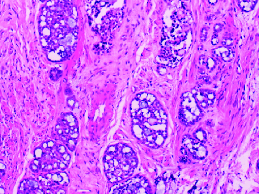
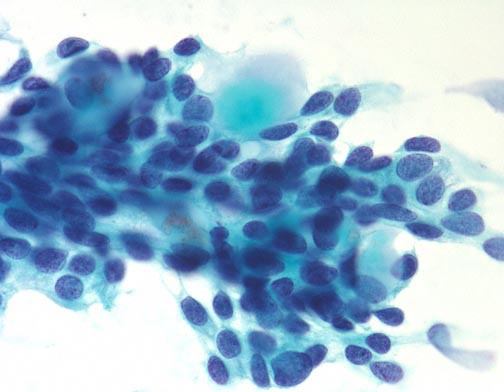
A fine needle biopsy or surgical removal of part or all of the tumour are two options for doing the biopsy. Fine needle aspiration, or FNA, is another name for a fine needle biopsy. A tiny needle is used to retrieve fluid and cells from the questionable location during this process. Adenoid cystic carcinoma tumours have a specific architecture in which epithelial cell bundles encircle and/or invade ducts or glandular structures within the organ. Adenoid cystic carcinoma is frequently diagnosed after the surgical excision of a tumour that was formerly deemed to be benign.
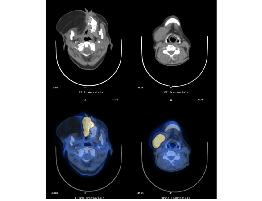
Imaging tests are performed. Before surgery, imaging procedures such as magnetic resonance imaging (MRI) or computed tomography (CT) scan can assist doctors in seeing the size and position of the tumour. If the tumour has migrated to other parts of the body, a positron emission tomography (PET) scan may be employed.


Your doctor will go through all of the results with you after the diagnostic tests are completed. These data can assist the doctor in describing cancer if the diagnosis is cancer. This is referred to as staging.
Enhance Strength & Mobility in Your Journey
For personalized guidance on cancer treatments and complementary therapies, consult our experts atZenOnco.ioor call+91 9930709000
Reference:
 Link Copied


Nurture Hope & Healing
with ZenOnco
On Google play India


Nurture Hope & Healing
with ZenOnco
On Google play India


